These articles are now archived and will no longer be updated.
Gut hormone blocks brain cell formation and is linked to Parkinson’s dementia, Swansea research reveals
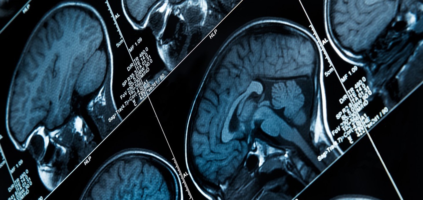
Brain scans: the research team focused on the gut hormone acyl-ghrelin (AG), which is known to promote brain cell formation
A gut hormone, ghrelin, is a key regulator of new nerve cells in the adult brain, a Swansea-led research team has discovered. It could help pave the way for new drugs to treat dementia in patients with Parkinson’s Disease.
Blood-borne factors such as hormones regulate the process of brain cell formation – known as neurogenesis - and cognition in adult mammals.
The research team focused on the gut hormone acyl-ghrelin (AG), which is known to promote brain cell formation. A structure change to the hormone results in two distinct forms: AG and unacylated-ghrelin (UAG).
The team, led by Dr Jeff Davies of Swansea University Medical School, studied both AG and UAG to examine their respective influences over brain cell formation.
This research is relevant to Parkinson’s as a large proportion of those with the disease experience dementia, which is linked to a loss of new nerve cells in the brain. This loss leads to a reduction in nerve cell connectivity, which plays a vital role in regulating memory function.
The team’s key overall findings were:
• the UAG form of ghrelin reduces nerve cell formation and impairs memory
• Individuals diagnosed with Parkinson’s disease dementia have a reduced AG:UAG ratio in their blood
Dr Jeff Davies of Swansea University Medical School, lead researcher, said:
“Our work highlights the crucial role of ghrelin as a regulator of new nerve cells in the adult brain, and the damaging effect of the UAG form specifically.
This hormone represents an important target for new drug research, which could lead ultimately to better treatment for people with Parkinson’s.
Our findings show that the AG:UAG ratio could also serve as a biomarker, allowing earlier identification of dementia in people with Parkinson’s disease.”
The team included collaborators from Newcastle University (UK) and Monash University (Australia). They examined the role of AG and UAG in the brain, and also compared blood collected from Parkinson’s disease patients diagnosed with dementia with cognitively intact PD patients and a control group.
They found:
• Higher levels of UAG, using both pharmacological and genetic methods, reduced hippocampal neurogenesis and brain plasticity.
• AG helped reverse spatial memory impairments
• UAG blocks the process of brain cell formation prompted by AG
• The Parkinson’s patients with dementia were the only one of the three patient groups examined to show a reduced AG:UAG ratio in their blood.
The research was published in Cell Reports Medicine.